
The word “mosaic” can appear on many kinds of genetic report: a pregnancy blood test, a chorionic villus sampling result, an embryo report from your IVF clinic, your child’s chromosome test, or even an adult cancer gene panel. It is rarely explained well. Many families leave the clinic unsure whether it is good news, bad news or something in between.
The honest answer is that mosaicism is a description, not a diagnosis. It tells you that not every cell in the body (or the embryo, or the placenta) carries the same genetic makeup. What that means for health depends on which genetic change is involved, how many cells carry it, and, most importantly, which parts of the body those cells are in.
This guide explains genetic mosaicism in plain language. It covers the different types, how it is tested, what it means in pregnancy and IVF, how it affects children and adults, and what it means for brothers, sisters and future pregnancies.
Quick answer
Genetic mosaicism means a person has two or more groups of cells with different genetic makeups, even though all their cells came from a single fertilised egg. It happens when a genetic change occurs after conception, during early cell division, so only the cells that come from the affected cell carry it. Its effect on health can range from none at all to the full features of a genetic condition.
What Is Genetic Mosaicism?
Every person begins as a single cell. That cell copies its DNA and divides, again and again, until it becomes the trillions of cells that make up the body. Each copy is usually perfect. Occasionally, though, a small error happens during one of these divisions. A chromosome may be gained or lost, or a single letter of the genetic code may change.
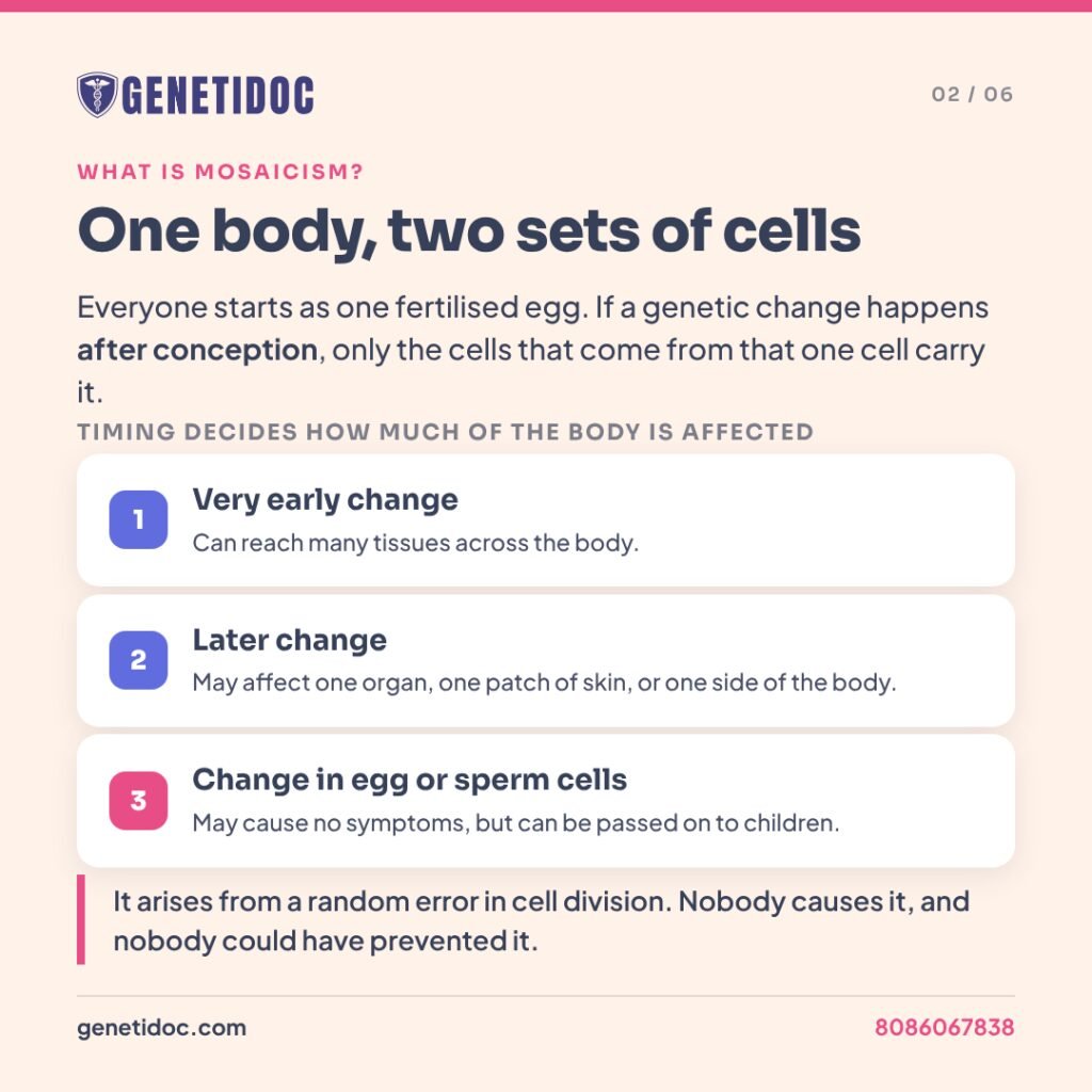
When that happens, every cell that later comes from the changed cell carries the same difference. All the other cells stay unchanged. The result is a body made of two “populations” of cells, like a mosaic floor made of two colours of tile. That is where the name comes from.
How much of the body is affected depends mainly on timing:
- A very early change, in the first few cell divisions, can end up in many tissues across the body.
- A later change may affect only one organ, one patch of skin, or one side of the body.
- A change in the cells that become eggs or sperm may cause no symptoms at all, but can be passed on to children.
Mosaicism is more common than doctors once thought. Older tests looked at only a small number of cells, so low levels of mosaicism were often missed. Modern techniques such as next-generation sequencing, which reads millions of DNA fragments at once, now detect it far more often.
The Main Types of Genetic Mosaicism
- Chromosomal mosaicism. Some cells have an extra or missing chromosome, or a missing or extra piece of one, while other cells are typical. Mosaic Down syndrome and mosaic Turner syndrome are well-known examples.
- Somatic mosaicism. A genetic change is present in some of the body’s cells but not in the eggs or sperm. It can cause patchy or one-sided features, such as overgrowth of one limb or streaks of lighter or darker skin.
- Germline (gonadal) mosaicism. The change is present in some egg or sperm cells. The parent’s blood test is often completely normal, yet more than one child can be affected.
- Confined placental mosaicism. The placenta carries a chromosome change, but the baby usually does not. This matters because pregnancy screening tests mostly read DNA from the placenta.
- Embryo mosaicism. An IVF embryo biopsy shows a mix of cells with typical and atypical chromosome numbers. This is reported on preimplantation genetic testing for aneuploidy (PGT-A), the test that checks embryos for missing or extra chromosomes.
Conditions Where Mosaicism Is Commonly Seen
Chromosomal conditions
- Mosaic Down syndrome accounts for about 2 percent of all people with Down syndrome. Some cells have three copies of chromosome 21 and others have the usual two.
- Mosaic Turner syndrome is common among girls and women with Turner syndrome. Some cells have one X chromosome and others carry a second sex chromosome.
- Pallister-Killian syndrome is caused by extra material from chromosome 12. It is often absent from blood and found only in skin or cheek cells.
- Mosaic trisomy 8 and mosaic trisomy 9, conditions that would usually not be survivable in the full, non-mosaic form.
Single-gene conditions
- PIK3CA-related overgrowth, where one part of the body grows larger than the rest.
- McCune-Albright syndrome (GNAS gene), which can affect bones, skin pigmentation and hormone glands.
- Sturge-Weber syndrome (GNAQ gene), linked to a facial port-wine birthmark and changes in the brain and eye.
- Segmental neurofibromatosis type 1 (NF1 gene), where features appear in only one region of the body.
- Pigmentary mosaicism, where swirls or streaks of lighter or darker skin follow faint lines on the skin called Blaschko lines. These lines trace the paths cells took as the embryo developed.
Signs That May Point Towards Mosaicism
Doctors may suspect mosaicism when they notice:
- One side of the body, one limb or one area growing larger than the other.
- Streaks, swirls or patches of skin colour following Blaschko lines.
- Features of a known genetic condition that are milder, patchier or less complete than usual.
- A strong clinical suspicion of a genetic condition, but a normal blood test.
- Two or more children with the same condition when both parents’ tests are normal.
- A pregnancy screening result that does not match ultrasound findings or later testing.
One published example shows why this matters. A 39-year-old woman with lines of darker skin on her arms and legs had a large duplication of chromosome 3. It was found only when DNA from a skin biopsy was tested (Martinez-Falero and colleagues, Clinical and Experimental Dermatology, 2022).
How Is Genetic Mosaicism Tested?
Why the sample matters as much as the test
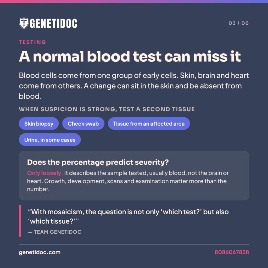
Blood is the easiest sample to collect, so most genetic tests use it. But blood cells come from just one group of early cells. Skin, brain, heart and other organs come from different groups. A change that arose after conception may be present in the skin or brain and completely absent in the blood.
This is why a normal blood result does not always rule out mosaicism. When the suspicion is strong, testing a second tissue is often the key step. Options include:
- A small skin biopsy from both the affected and unaffected areas, sometimes grown in the laboratory as cultured skin cells (fibroblasts).
- A cheek (buccal) swab.
- Tissue from an affected area, for example tissue removed during surgery for overgrowth.
- Urine, in some situations.
“A normal report means no reportable finding in the sample and the test that was used. It is not a guarantee,” says Team Genetidoc. “With mosaicism, the question is not only ‘which test?’ but also ‘which tissue?’ If the clinical picture still points strongly to a genetic cause, we look at a second tissue before concluding anything.”
Which test is used
| Test | What it detects | Mosaicism limits |
| Karyotype (chromosome count under a microscope) | Extra, missing or rearranged chromosomes | A standard 20-cell count reliably detects only about 14 percent mosaicism or more. Counting 30 to 50 cells improves this to about 6 to 10 percent. |
| Chromosomal microarray | Small missing or extra pieces of chromosomes | Usually detects mosaicism above roughly 10 to 20 percent, depending on the platform |
| Fluorescence in situ hybridisation (FISH) | A specific chromosome, checked in hundreds of cells | Useful for confirming a suspected mosaic chromosome change |
| Deep next-generation sequencing | Single-gene changes present in a small share of cells | Can detect low-level mosaicism, often from affected tissue rather than blood |
A routine karyotype usually takes about three to four weeks. Tissue-based or deep-sequencing tests can take longer because the sample may need to be grown in the laboratory first.
Does the percentage on the report predict severity?
This is one of the most common questions families ask, and the honest answer is: only loosely. The percentage on a report describes the sample that was tested, usually blood. It may be very different in the brain, heart or other organs, and those organs are what shape a person’s health. A child with a low percentage in blood can have significant features, and a child with a higher percentage can do very well.
Counseling therefore focuses on the person in front of us: their growth, development, scans and examination findings. The number on the page is only one part of that picture.
Mosaicism in Pregnancy: Non-Invasive Prenatal Testing and Chorionic Villus Sampling Results
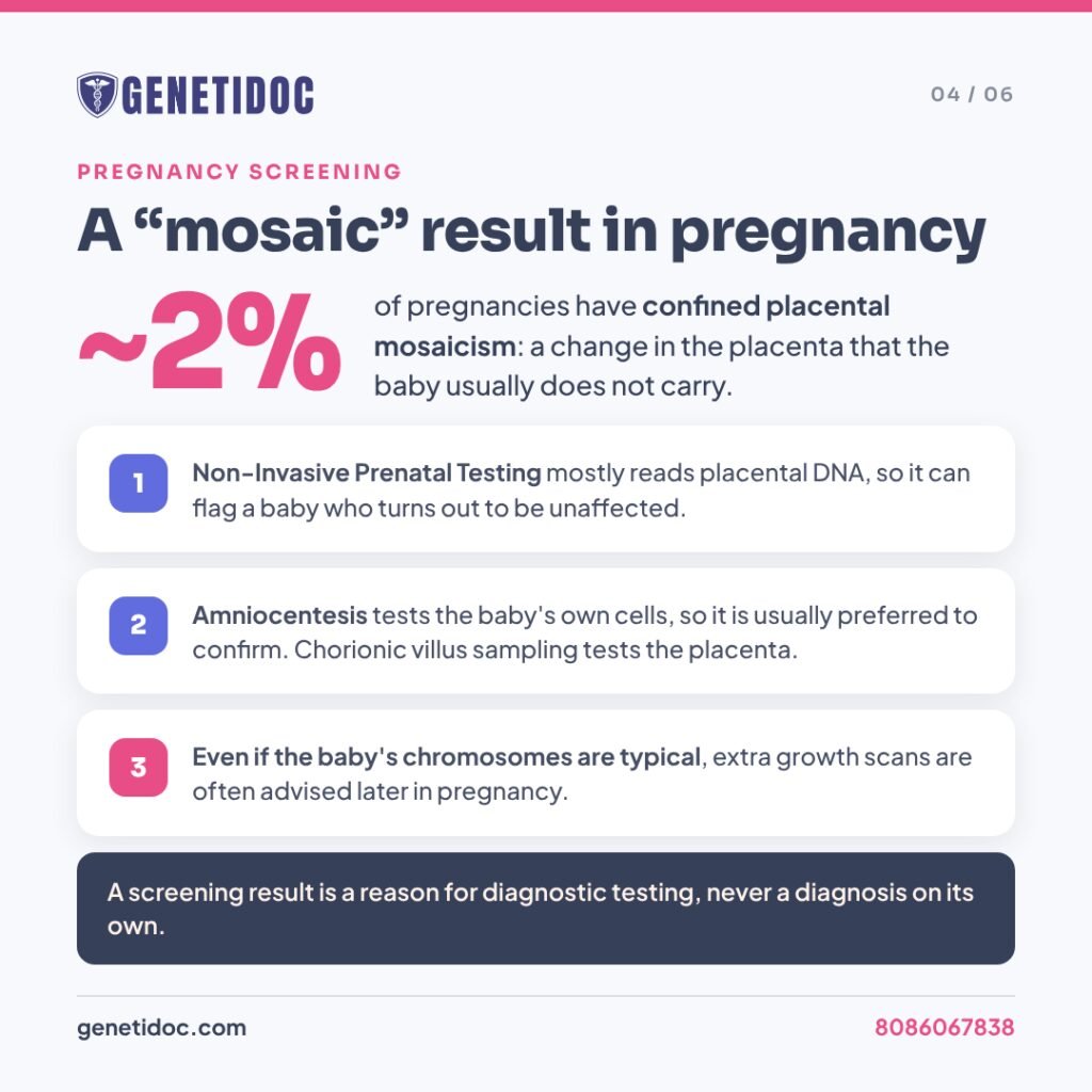
Non-Invasive Prenatal Testing (NIPT) analyses small fragments of DNA in the mother’s blood. Most of this DNA comes from the placenta, not directly from the baby. Usually the placenta and baby share the same chromosomes, but not always.
In confined placental mosaicism, a chromosome change is present in the placenta and the baby is usually chromosomally typical. It affects around 2 percent of pregnancies (Eggenhuizen and colleagues, Human Reproduction Update, 2021). It is one of the main reasons NIPT can give a high-risk result for a baby who turns out to be unaffected.
“Placental mosaicism is one of the limitations we discuss with every couple before NIPT, along with a vanishing twin and low fetal fraction,” explains Team Genetidoc. “NIPT is a screening test. A high-risk or mosaic result is a reason for diagnostic testing, never a diagnosis on its own.”
Why amniocentesis is usually preferred to confirm a mosaic result
Chorionic villus sampling takes a small sample of the placenta, so it can show the same placental mosaicism that NIPT picked up. Amniocentesis tests cells from the fluid around the baby, which come from the baby itself. So when NIPT or chorionic villus sampling suggests mosaicism, amniocentesis is usually recommended. It is followed by karyotyping or chromosomal microarray analysis.
When the baby’s chromosomes are normal
Even when confined placental mosaicism is confirmed and the baby’s chromosomes are typical, the pregnancy still needs attention. A placenta with abnormal cells may not work as efficiently, and this is linked to slower fetal growth. Trisomy 16 in the placenta carries the highest risk. Closer growth scans and placental assessment later in pregnancy are commonly advised.
For some chromosomes, an additional test is recommended. Guidance from the American College of Medical Genetics and Genomics (ACMG, 2020) advises testing for uniparental disomy when mosaic trisomy involves chromosomes 6, 7, 11, 14, 15 or 20. Uniparental disomy means the baby inherited both copies of a chromosome from one parent. For these particular chromosomes, which parent a copy came from affects how certain genes work.
Illustrative example
A woman has NIPT at 11 weeks, and it shows a high-risk result for trisomy 16. Her ultrasound looks normal. Amniocentesis at 16 weeks shows typical chromosomes in the baby, which points to confined placental mosaicism. Uniparental disomy testing is not needed because chromosome 16 is not one of the listed imprinted chromosomes. However, her obstetrician arranges extra growth scans in the third trimester, because placental mosaicism for chromosome 16 is linked to slower fetal growth.
Genetidoc offers NIPT and the counseling around it. Invasive procedures such as amniocentesis and chorionic villus sampling are performed by your obstetrician or fetal medicine specialist. Our team helps you understand what the results mean.
Mosaic Embryos After IVF
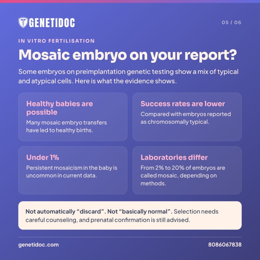
During IVF with PGT-A, a few cells are taken from the outer layer of a five- or six-day-old embryo (the trophectoderm, which later forms the placenta). If the result falls between clearly normal and clearly abnormal, the embryo is often reported as “mosaic”. Studies commonly use this label when roughly 20 to 80 percent of the biopsied cells appear abnormal.
What the evidence shows
- Healthy babies are possible. Many mosaic embryo transfers have resulted in healthy births.
- Success rates are generally lower than for embryos reported as euploid, meaning chromosomally typical.
- Persistent mosaicism in the baby is uncommon. Current data suggest it is under 1 percent, though it has been reported.
- Laboratories differ. The share of embryos called mosaic varies from about 2 percent to 20 percent between PGT laboratories, depending on their methods and cut-offs.
How mosaic embryos are prioritised
The Preimplantation Genetic Diagnosis International Society (PGDIS) has published position statements on mosaic embryo transfer. Generally:
- Euploid embryos are transferred first.
- Low-level mosaic embryos are preferred over high-level ones.
- Changes affecting part of a chromosome (segmental) are generally preferred over changes affecting a whole chromosome.
- Chromosomes linked to trisomies that can survive to birth (13, 18, 21) or to uniparental disomy are approached more cautiously.
The American Society for Reproductive Medicine (ASRM, 2023) notes that there is not yet enough data for firm, evidence-based recommendations on prenatal testing after mosaic embryo transfer. Prenatal diagnosis is generally offered, usually amniocentesis rather than chorionic villus sampling.
A published case shows why this matters. A couple with only one complex mosaic embryo chose to transfer it after counseling. Amniocentesis later confirmed mosaic trisomy 7 at about 20 percent, and serial scans showed no structural abnormalities (Pham and Phan, 2026).
Two misconceptions to avoid
- “Mosaic means abnormal, so it should be discarded.” Not necessarily. For couples with no euploid embryos, a carefully chosen mosaic embryo may still be a reasonable option.
- “Mosaic is basically normal.” Also not true. Outcomes differ from those of euploid embryos, and confirmation in pregnancy is still advised.
Whether to transfer a mosaic embryo is the couple’s decision. Our role is to explain the evidence clearly so the couple can make it with full information.
Mosaicism in Children: What a Diagnosis Changes
For parents, a mosaic diagnosis can feel confusing. Your child has a named condition, but perhaps not “fully”. In practice, a diagnosis still changes a lot:
- It ends the search. Many families have been through years of tests before mosaicism is identified through the right tissue.
- It guides screening. A child with mosaic Down syndrome still needs routine heart, thyroid, hearing, vision and developmental checks. These follow the same guidelines as for full Down syndrome.
- It flags specific risks. In mosaic Turner syndrome, testing for Y chromosome material is important. If it is present, there is a higher risk of a tumour in the ovaries, which can be prevented.
- It can open treatment options. For some mosaic overgrowth conditions caused by changes in genes such as PIK3CA, targeted medicines that act on the affected cell pathway are now being used or studied.
- It informs family planning, as explained below.
Children with mosaicism may, on average, have milder features than those with the full form of the same condition. But the range is wide and overlapping. “Mosaic” should never be read as an automatic promise of “mild”.
Mosaicism in Adults and Cancer Gene Testing
Mosaicism can also be found in adults. Sometimes it shows up after a hereditary cancer panel. Sometimes a parent turns out to have mild features only after their child is diagnosed with the full form of a condition.
On cancer panels, a genetic change found in only a small share of blood cells needs careful interpretation. It may represent:
- True mosaicism, present in some cells throughout the body.
- Clonal hematopoiesis, an age-related change limited to blood-forming cells. It is not inherited and not passed on, and it is seen most often in genes such as TP53, CHEK2 and ATM.
- An undiagnosed blood cancer.
- DNA released from a tumour into the bloodstream.
“When we see a low-level TP53 change in blood, we don’t label it inherited straight away,” says Team Genetidoc. “It can reflect clonal hematopoiesis, mosaicism, a blood cancer or tumour DNA. Confirming it on a non-blood tissue, such as cultured skin cells, is what tells us whether the family needs testing.”
When true mosaicism is confirmed, cancer screening usually follows the guidelines for the full condition, adjusted to the person’s own history. In segmental neurofibromatosis type 1, for example, follow-up focuses on the affected region. Management is always built around personal and family history, not the laboratory number alone.
Is Mosaicism Inherited? What It Means for Your Family
If a person has mosaicism
Mosaicism itself usually arises after conception, so it is generally not inherited from a parent. The question is whether the change is present in that person’s eggs or sperm. If it is, a child can inherit it, and the child will carry it in every cell. In other words, a mildly affected parent can have a child with the full, non-mosaic form of the condition. Many adults only discover their own mosaicism this way.
If your child’s condition was “de novo” and your tests were normal
Parents are often told that their child’s genetic change is de novo, meaning new in the child and not found in either parent’s blood. This is reassuring, but not a complete guarantee. A parent can carry the change in some egg or sperm cells, which is called germline or gonadal mosaicism, while their blood test is entirely normal.
For this reason, the chance of the same condition happening again is small but not zero. For many conditions it is often quoted at around 1 percent. It is higher for some conditions, such as Duchenne muscular dystrophy, where germline mosaicism in mothers with normal carrier tests is well documented. Your exact figure depends on the condition and should be discussed in counseling.
Illustrative example
A couple’s first child has a dominant bone condition caused by a de novo change. Both parents test negative. They are counseled that the chance of recurrence is low but not zero, because of possible germline mosaicism. In their next pregnancy, they choose prenatal testing for the specific family change for reassurance.
Options for future pregnancies
- Prenatal diagnosis for the specific family change in a future pregnancy.
- Preimplantation genetic testing during IVF, which tests embryos for the known change before transfer.
- Testing other family members when a parent’s own mosaicism is confirmed.
“Parents often carry silent guilt, wondering if they ‘passed something on’,” says Team Genetidoc. “Mosaicism arises from a random error in cell division. Nobody causes it, and nobody could have prevented it. Part of counseling is making sure parents hear that clearly.”
When Testing for Mosaicism Is Useful, and When It May Not Be
Testing is useful when
- A child has patchy skin changes, asymmetry or overgrowth.
- A genetic condition is strongly suspected but blood testing is normal.
- A pregnancy screening result needs confirming or clarifying.
- An IVF couple needs help understanding a mosaic embryo report.
- More than one child in a family is affected despite normal parental tests.
- A low-level change appears on a cancer panel.
Testing may add little when
- The result would not change care, screening or family planning.
- Repeated testing of the same tissue is unlikely to show anything new. A different tissue is usually the better step.
- The test is chosen without a clinical question in mind. Mosaicism testing works best when it is guided by examination and history.
Why Expert Interpretation Matters So Much With Mosaicism
Few areas of genetics depend on interpretation as heavily as mosaicism. The same laboratory result can mean very different things depending on the sample, the test’s sensitivity, the chromosome or gene involved, and the person’s own features. Common problems when results are not interpreted carefully include:
- Mosaic embryos discarded without the couple understanding their options.
- Placental mosaicism on NIPT mistaken for a confirmed problem in the baby.
- Low-level mosaicism missed because too few cells were counted, or the wrong tissue was tested.
- Age-related blood changes mislabelled as an inherited cancer gene, causing unnecessary worry for relatives.
Choosing an accredited laboratory, the right test and the right tissue matters. So does having a clinical geneticist and genetic counselor who connect the result to the person. Genetidoc’s NABL and CAP accredited team builds genetic counseling into every result. After consultation, families receive an individualised written summary they can refer back to and share with their doctors.
What to Do If Your Report Mentions Mosaicism
- Don’t panic, and don’t assume it means “mild”. Mosaicism is a description, not a prognosis.
- Check which sample was tested. Blood, placenta, embryo or skin results each mean something different.
- Confirm pregnancy results with diagnostic testing. This is usually amniocentesis.
- Don’t base decisions on the percentage alone. Clinical features and scans matter more.
- Keep your reports and raw data. They help with future review, family testing and pregnancy planning.
- See a genetic counselor before major decisions, such as embryo transfer or continuing or ending a pregnancy.
- Return for review if new symptoms appear, or before planning another pregnancy.
Families can also read about others’ experiences and ask questions on the Genetidoc Rare Disease Forum, including the Non-Invasive Prenatal Testing and Preimplantation Genetic Testing discussions.
Book your genetic consultation
Frequently Asked Questions
What does mosaicism mean in simple terms?
It means some cells in the body have a different genetic makeup from others. This happens because a genetic change occurred after conception, during early cell division.
Is genetic mosaicism inherited?
Usually not. It typically arises after conception. However, if the change is present in a person’s eggs or sperm, it can be passed to a child, who may then have the full form of the condition.
Is mosaic Down syndrome milder than Down syndrome?
On average, features may be milder, but the range is wide and overlaps with full Down syndrome. Children with mosaic Down syndrome should follow the same health screening schedule.
Does a higher percentage of mosaicism mean a more severe condition?
Not reliably. The percentage reflects only the tissue tested, usually blood, and may differ in organs like the brain or heart. Clinical features matter more than the number.
Can a blood test miss mosaicism?
Yes. A change may be present in skin or other tissues but absent from blood. Testing a second tissue, such as a skin biopsy or cheek swab, is often needed.
What is confined placental mosaicism?
It is a chromosome change found in the placenta but usually not in the baby. It affects around 2 percent of pregnancies and is a known cause of false-positive NIPT results.
My NIPT shows a mosaic result. What should I do next?
Speak to a genetic counselor and your obstetrician. Diagnostic testing, usually amniocentesis, is recommended to check the baby’s own chromosomes before any decision is made.
Can a mosaic embryo result in a healthy baby?
Yes. Many mosaic embryo transfers have led to healthy births, although success rates are generally lower than with euploid embryos. Prenatal diagnostic testing is usually advised.
What is germline mosaicism?
It is when a genetic change is present in some of a parent’s egg or sperm cells but not in their blood. It explains why a “de novo” condition can occasionally happen again in a family.
If my child’s condition is de novo, can it happen again?
The chance is small but not zero, because of possible germline mosaicism. It is often around 1 percent and higher for some conditions. Prenatal testing can be offered in future pregnancies.
Can mosaicism be prevented or cured?
It cannot be prevented, because it results from random errors in cell division. Treatment focuses on the specific condition, and targeted medicines are emerging for some mosaic overgrowth conditions.
Can a low-level variant on my cancer gene test be mosaicism?
It can, but it may also reflect age-related blood changes or other causes. Confirmation on a non-blood tissue helps decide whether it is inherited and whether relatives need testing.
Key Takeaways
- Genetic mosaicism means having two or more groups of cells with different genetic makeups. It is a description, not a diagnosis.
- Its effects depend on the genetic change, the share of cells affected, and which tissues carry it.
- A normal blood test does not always rule it out, and testing a second tissue is often key.
- Mosaic NIPT or chorionic villus sampling results need confirmation, usually by amniocentesis.
- Mosaic embryos can lead to healthy babies, but they need careful selection and prenatal confirmation.
- Even “de novo” conditions carry a small recurrence risk because of germline mosaicism.
- Expert interpretation and genetic counseling are what turn a mosaic result into a clear plan.
Has a report — yours, your baby’s, your embryo’s or your child’s — mentioned mosaicism?
Our clinical geneticists can review the report, explain what the sample and percentage really mean, and guide next steps, from confirmatory testing to planning a future pregnancy. Consultations are available online across India and in person in Thiruvananthapuram. Book a mosaicism report review with a Genetidoc clinical geneticist, or call 8086067838.
References
- Eggenhuizen GM, Go A, Koster MPH, et al. Confined placental mosaicism and the association with pregnancy outcome and fetal growth: a review of the literature. Human Reproduction Update. 2021;27(5):885–903. doi:10.1093/humupd/dmab009
- Cram DS, Leigh D, Handyside A, et al. PGDIS position statement on the transfer of mosaic embryos 2019. Reproductive BioMedicine Online. 2019;39(4):e1–e4
- American College of Medical Genetics and Genomics. Clinical utility of testing for uniparental disomy (points to consider), Genetics in Medicine, 2020
- American Society for Reproductive Medicine Practice Committee. Clinical management of mosaic results of preimplantation genetic testing for aneuploidy (PGT-A) of blastocysts: a committee opinion, Fertility and Sterility, 2023
- Greco E, Minasi MG, Fiorentino F. Healthy babies after intrauterine transfer of mosaic aneuploid blastocysts. New England Journal of Medicine. 2015;373(21):2089–2090
- Sait H. Mosaicism in clinical genetics: counselling challenges and diagnostic dilemmas. Genetic Clinics (Indian Academy of Medical Genetics). 2024;17(3):3–14
- Has C. Genetic testing in mosaicism. JEADV Clinical Practice. 2025;4(3):664–668. doi:10.1002/jvc2.70073
- Waldman L, Mahon SM. Mosaicism: implications for genetic counseling and medical management. Clinical Journal of Oncology Nursing. 2017;21(4):428–432
- Martinez-Falero BS, et al. Pigmentary anomaly caused by mosaic 3q22.2q29 duplication. Clinical and Experimental Dermatology. 2022;47(12):2342–2345
- Pham TTD, Phan TM. A case report of prenatal-confirmed trisomy 7 mosaicism after complex mosaic embryo transfer. Tạp chí Y học Cộng đồng. 2026;67(CĐ9)