
Quick answer
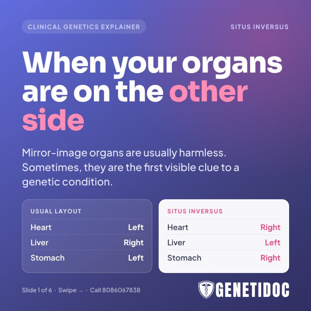
Situs inversus is a rare condition, present from birth, in which the organs of the chest and abdomen sit in a mirror-image position. The heart is on the right, the liver on the left, and the stomach on the right. On its own, complete mirror-image arrangement usually causes no health problems. However, it can sometimes be a sign of an inherited condition called primary ciliary dyskinesia, or part of a more complex pattern called heterotaxy. These need proper medical and genetic evaluation.
Many people first hear the words “situs inversus” in a surprising way. A radiologist mentions it during a routine chest X-ray. A doctor notices that the heartbeat is louder on the right. Or an expectant mother is told during her anomaly scan that her baby’s heart or stomach is “on the other side.”
The first reaction is usually worry. The second is a flood of online searches, many of which simply call situs inversus a harmless medical curiosity.
The truth sits in between. For many people, mirror-image organs are just a different arrangement that never causes trouble. For others, it is the first visible clue to a genetic condition that affects the lungs, sinuses, ears, fertility or heart. Knowing which group you or your child belongs to makes a real difference.
This guide explains what situs inversus is, why it happens, when it matters, and how genetic evaluation helps families in India get clear answers.
“Finding organs on the opposite side is not a diagnosis in itself. It is a question. Our job is to find out whether it is simply a different arrangement or a sign of something the family needs to know about.”
— Dr. Roshan Daniel, Clinical Geneticist, Genetidoc
What Is Situs Inversus?
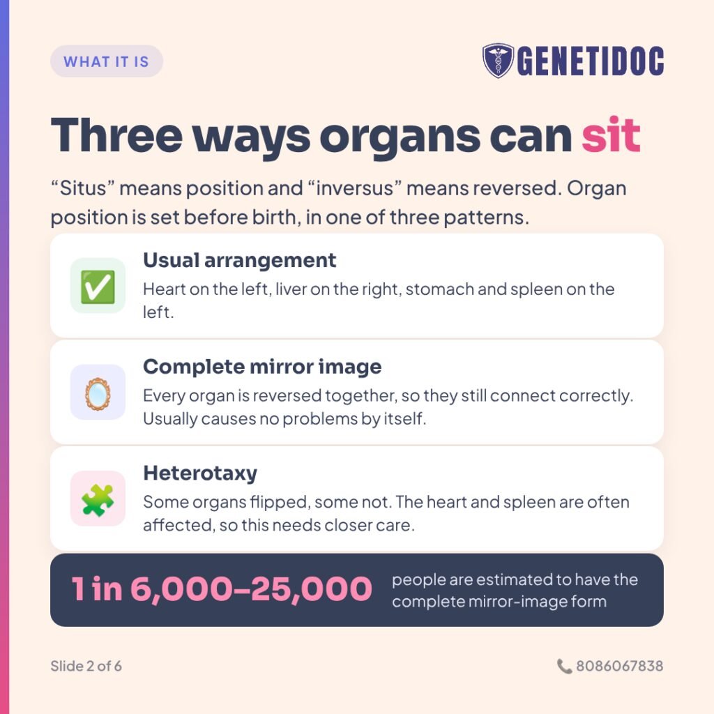
“Situs” is a Latin word meaning “position.” “Inversus” means “reversed.” So situs inversus simply means the organs are in a reversed position.
In most people, the heart points to the left, the liver sits on the right side of the upper abdomen, and the stomach and spleen sit on the left. Doctors call this normal arrangement situs solitus.
In situs inversus totalis (complete situs inversus), every one of these organs is flipped, like a reflection in a mirror. Because the whole arrangement is reversed together, the organs still connect to each other correctly. This is why many people with complete situs inversus live completely normal lives and only discover it by chance.
Situs inversus totalis is rare. It is estimated to affect roughly 1 in 6,000 to 1 in 25,000 people.
The three main patterns of organ arrangement
- Situs solitus (normal arrangement): Heart on the left, liver on the right, stomach and spleen on the left.
- Situs inversus totalis (complete mirror image): All chest and abdominal organs are reversed. Usually causes no problems by itself.
- Situs ambiguus, also called heterotaxy: The organs are neither normal nor a perfect mirror image. Some are flipped and some are not, and organs like the heart and spleen are often structurally abnormal. This pattern is much more likely to cause serious health problems.
What is dextrocardia?
Dextrocardia means the heart is on the right side of the chest. It is part of situs inversus totalis, but it can also occur on its own while the abdominal organs stay in their normal positions. Isolated dextrocardia (dextrocardia without the rest of the organs being mirror-reversed) is more often linked to heart defects. Anyone found to have it should have a detailed heart evaluation, including an echocardiogram (an ultrasound scan of the heart).
Why Do Organs End Up on the Wrong Side?
The answer lies in a tiny structure most people have never heard of: the cilium (plural: cilia).
Cilia are microscopic, hair-like structures on the surface of many cells. Some cilia can move and beat in a coordinated rhythm. In the airways, they work like a moving carpet, sweeping mucus, dust and germs up and out of the lungs.
Very early in pregnancy, in the first few weeks after conception, a special group of moving cilia in the embryo beat in one direction. This creates a gentle flow of fluid towards the left side. That flow switches on a chain of “left-side” genetic signals. These signals tell the developing heart, lungs, liver, stomach and spleen which way to turn and where to settle.
If those cilia do not move properly, the embryo loses its sense of left and right. The organs then settle on one side or the other largely by chance. This is why about half of people with non-moving cilia have normal organ positions and about half have mirror-image organs, even among brothers and sisters in the same family.
Problems in the genes that send the left-right signals can also disturb this process, even when the cilia themselves are healthy. Some of these genes are listed later in this article.
Is Situs Inversus Dangerous?
Short answer: Complete situs inversus by itself is usually not dangerous, and life expectancy is normal. The real question is whether it is isolated, or part of primary ciliary dyskinesia or heterotaxy. Those conditions need specialist care.
In practice, people with mirror-image organs fall into three broad groups.
1. Isolated situs inversus totalis
The organs are a perfect mirror image, the heart is structurally normal, and there are no chest, sinus, ear or fertility problems. Most of these people stay healthy throughout life. A small minority have heart defects, which is why a heart check is still sensible.
2. Situs inversus with primary ciliary dyskinesia (Kartagener syndrome)
The organs are mirror-reversed because the cilia do not move properly. The same faulty cilia also cause lifelong lung, sinus and ear problems, and often infertility. Mirror-image organs combined with primary ciliary dyskinesia is traditionally called Kartagener syndrome, after the doctor who described it in 1933.
3. Heterotaxy (situs ambiguus)
The organs are jumbled rather than neatly mirrored. This group has the highest risk of complex heart defects, spleen problems and twisting of the bowel. Most children with heterotaxy need care from a pediatric cardiologist from birth.
“The position of the organs tells us very little on its own. The chest, the sinuses, the ears, the heart scan and the family history tell us the rest. That full picture is what decides whether a person needs testing, and which test.”
— Dr. Roshan Daniel, Clinical Geneticist, Genetidoc
Situs Inversus and Primary Ciliary Dyskinesia
Primary ciliary dyskinesia (PCD) is an inherited condition in which the moving cilia in the body do not beat properly. “Primary” means it is present from birth, “ciliary” refers to the cilia, and “dyskinesia” means abnormal movement.
Because cilia cannot clear mucus, it builds up in the airways, sinuses and middle ears. Over time, repeated infections can damage the airways and cause bronchiectasis, a condition in which the breathing tubes become permanently widened and scarred.
Large studies show that roughly half of people with PCD have situs inversus totalis. A smaller group, estimated at around 6 to 12 percent, have heterotaxy. This means two important things:
- Mirror-image organs can be a valuable early clue to PCD.
- Normal organ position does not rule PCD out. Around half of people with PCD have their organs in the usual place.
Worldwide, PCD is estimated to affect about 1 in 10,000 to 15,000 people. However, it is more common in communities where marriages between relatives are common. This is relevant for many Indian families, as discussed below.
Warning signs that situs inversus may be part of PCD
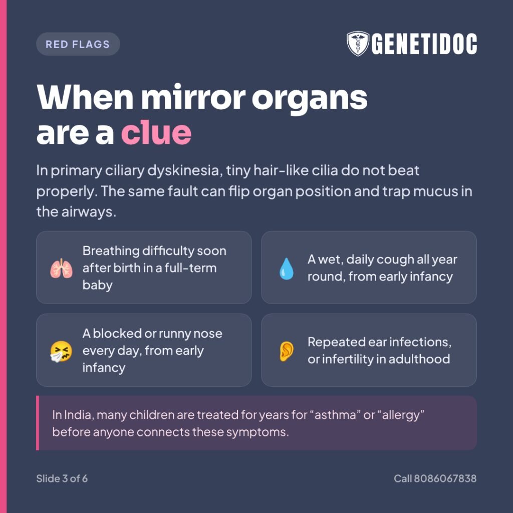
The American Thoracic Society highlights four key features of PCD. A person with mirror-image organs who has even one of the other three should be evaluated:
- Breathing difficulty soon after birth in a baby born at full term, without an obvious cause, often needing oxygen for days.
- A wet, daily cough all year round, starting in early infancy, not just during colds.
- A blocked or runny nose every day, all year round, starting in early infancy.
- Organs in an abnormal position, such as situs inversus or heterotaxy.
Other clues include repeated ear infections, persistent fluid behind the eardrums (“glue ear”), hearing problems in childhood, chronic sinusitis, recurrent pneumonia, and bronchiectasis found on a CT scan. In adults, a clue may be infertility in men or difficulty conceiving or ectopic pregnancy in women.
In India, many children with PCD are treated for years for “asthma,” “allergy,” “bronchitis” or “frequent colds” before anyone connects the symptoms. Mirror-image organs on a chest X-ray, combined with a lifelong wet cough, should always raise the question of PCD.
Heterotaxy: When Organs Are Neither Normal nor Mirrored
Heterotaxy syndrome (from Greek words meaning “different arrangement”) affects about 1 in 10,000 people and accounts for about 3 percent of congenital heart defects. In heterotaxy, the body behaves as if it has two right sides or two left sides. Doctors call this isomerism.
Right isomerism (sometimes called asplenia syndrome)
- Often complex, serious heart defects
- Spleen may be missing (asplenia), which raises the risk of severe infections
- Bowel may be abnormally rotated (intestinal malrotation), which can cause dangerous twisting
Left isomerism (sometimes called polysplenia syndrome)
- Multiple small spleens, which may not work normally
- Heart defects, often less severe than in right isomerism
- Problems with the heart’s electrical system, including slow heart rhythm (heart block)
Heterotaxy varies greatly from person to person, even within one family. Some children need heart surgery in infancy. Others have milder forms that are found later in life.
Is Situs Inversus Genetic?
Short answer: Often, yes, but not always. When situs inversus is part of PCD or heterotaxy, a genetic cause can frequently be found. In truly isolated situs inversus with no other features, a cause is often not identified with current testing.
Genetics of primary ciliary dyskinesia
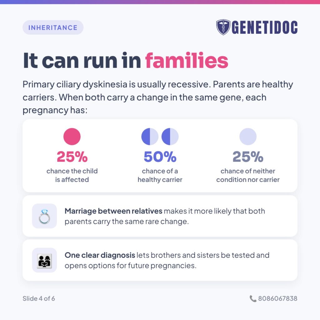
PCD is usually inherited in an autosomal recessive pattern. This means a child is affected only when they inherit a non-working copy of the same gene from both parents. The parents are usually healthy carriers. They each carry one non-working copy and one working copy, so they have no symptoms.
When both parents are carriers, each pregnancy has:
- A 1 in 4 (25 percent) chance of a child with PCD
- A 2 in 4 (50 percent) chance of a child who is a healthy carrier
- A 1 in 4 (25 percent) chance of a child who neither has PCD nor is a carrier
More than 50 genes are now linked to PCD, and the list keeps growing. Commonly involved genes worldwide include DNAH5, DNAH11, DNAI1, CCDC39 and CCDC40. A few rare forms are X-linked, meaning the gene is on the X chromosome and mainly affects boys.
What we know about PCD in India
Indian research on PCD genetics is still limited, but it is growing. In one Indian study of children with suspected PCD who had whole exome sequencing, the most common genes in confirmed cases were LRRC6, DNAH5, CCDC39 and HYDIN. Several of the genetic changes found had never been reported anywhere before. The researchers suggested that India may need gene panels designed for its own population.
This matters because a test designed mainly around Western patients may not be the best fit for an Indian patient. It is one reason why careful test selection and expert interpretation are so important.
India also has many communities where marriage between relatives (consanguinity) is a long-standing tradition. In such families, both parents are more likely to carry the same rare genetic change, and recessive conditions like PCD become more common. Studies from other highly consanguineous populations show that a family’s specific genetic change can often be found quickly with targeted testing once it is known in one relative.
“When parents learn their child has a recessive condition, especially in a marriage between relatives, guilt often comes first. We are very clear with families: nobody chooses the genes they carry. A diagnosis is not about blame. It is about protecting this child and planning for the next.”
— Dr. Roshan Daniel, Clinical Geneticist, Genetidoc
Genetics of heterotaxy
Heterotaxy is genetically complex. It can follow different inheritance patterns depending on the gene involved:
- Autosomal recessive, as with several cilia genes such as DNAH5 and DNAH11
- Autosomal dominant, where one changed copy is enough, as with some genes in the left-right signaling pathway such as NODAL and LEFTY2
- X-linked, most notably the ZIC3 gene, which mainly affects boys
No single gene explains most cases. ZIC3 accounts for only a small share of heterotaxy, and the signaling-pathway genes together explain perhaps 5 to 10 percent. Research has also found that missing or extra pieces of chromosomes, called copy number variants, are present in roughly one in five people with heterotaxy. Standard gene sequencing can miss these. A separate test called chromosomal microarray is designed to detect them.
When Is Genetic Testing Useful?
Genetic evaluation is generally worth discussing with a clinical geneticist if any of the following apply:
- Situs inversus or dextrocardia plus a long-standing wet cough, chronic sinus problems, repeated ear infections or bronchiectasis
- Breathing difficulty at birth in a full-term baby who also has abnormal organ position
- Heterotaxy, especially with a congenital heart defect or spleen abnormality
- Infertility in a man with poorly moving sperm, or repeated ectopic pregnancies, especially with any chest or sinus history
- A brother, sister or cousin already diagnosed with PCD or heterotaxy
- Parents who are blood relatives and have a child with organ position abnormalities
- A couple whose previous baby had heterotaxy or PCD and who are planning another pregnancy
- A pregnancy in which the anomaly scan shows abnormal organ position, especially with a heart defect
When Genetic Testing May Not Be Needed
Genetic testing is not automatically required for everyone with mirror-image organs. Testing may add little if:
- You are an adult with complete, perfectly mirrored situs inversus
- Your heart scan is normal
- You have no lifelong cough, sinus, ear or fertility problems
- No one in your family has a related condition
In this situation, a careful clinical review is often enough. Testing may still be worth discussing if you are planning a family and want to understand the risk for your children, or if new symptoms appear later.
The decision to test belongs to you. A genetic counselor’s role is to explain the options and their limits clearly, not to push you in either direction.
How Is Situs Inversus Evaluated? A Step-by-Step Approach
Step 1: Confirm the organ arrangement
Doctors use a chest X-ray, abdominal ultrasound and sometimes a CT or MRI scan to map exactly where each organ sits. They also check whether the spleen is present, absent or multiple.
Step 2: Check the heart
An echocardiogram looks at the structure of the heart. An electrocardiogram (ECG, a recording of the heart’s electrical activity) checks its rhythm.
Step 3: Take a detailed history
The doctor asks about breathing at birth, cough, sinuses, ears, hearing, fertility and pregnancy history. A three-generation family history is drawn, including whether the parents are related.
Step 4: Tests for PCD, where it is suspected
Several specialized tests can support a diagnosis of PCD:
- Nasal nitric oxide: Measures a gas that is usually very low in PCD. It is useful from around five years of age.
- High-speed video microscopy: Records how cilia from a nasal brushing actually beat.
- Transmission electron microscopy: Looks at the internal structure of cilia under a very powerful microscope.
- Immunofluorescence: Uses special stains to show whether key cilia proteins are present.
These tests are available only at a few specialized centers in India. They also have important limitations. Nasal nitric oxide can be normal in some genetic forms of PCD. Electron microscopy can look normal or be inconclusive in up to about a third of patients, because some genetic faults change how cilia move without changing how they look.
This is why genetic testing plays such an important role. American Thoracic Society guidelines accept two disease-causing changes in a known PCD gene as confirmation of the diagnosis.
Step 5: Choosing the right genetic test
The right test depends on the clinical picture:
- Targeted variant testing: If a relative already has a confirmed genetic diagnosis, testing for that specific change is quick and focused. Turnaround is usually around a week.
- PCD multi-gene panel: For suspected PCD, a panel examines many cilia-related genes at once. Turnaround is typically around 3 to 4 weeks.
- Heterotaxy or laterality panel: For heterotaxy, this covers both cilia genes and left-right signaling genes.
- Chromosomal microarray: Often added for heterotaxy with heart defects, to look for missing or extra chromosome pieces.
- Whole exome sequencing: Reads nearly all the protein-coding genes. It is useful when the picture is unusual or a panel is negative despite strong suspicion. It typically takes longer than a panel.
Costs vary depending on the type and scope of the test. Your genetic counselor can explain the options before you decide.
Why Interpretation Matters More Than the Report
A genetic report is only as useful as the interpretation behind it. In situs inversus, PCD and heterotaxy, several things can go wrong when testing is done without expert input.
The wrong test can miss the answer
A panel built for PCD may not include the left-right signaling genes involved in heterotaxy. Gene sequencing alone may miss a deleted chromosome piece that only a microarray would find. Some laboratories also do not look carefully for small deletions of single sections of a gene, which have been found in a meaningful share of PCD diagnoses.
“One change found” is not a diagnosis
Because PCD is usually recessive, finding a change in only one copy of a gene does not confirm the condition. It may mean the person is simply a carrier. Or the second change may be hiding where the test did not look. Expert review decides whether more testing is needed.
Uncertain results need careful handling
Many PCD genes are large, and reports often include a variant of uncertain significance. This is a genetic change that science cannot yet classify as harmful or harmless. It should not be used to make decisions about relatives or future pregnancies. Testing affected and unaffected family members can sometimes help clarify what the change means. Reviewing the result again after 6 months to a year can also help, as new research becomes available.
A negative result is not always the final word
A negative test means no reportable change was found in what was tested. It does not prove that a person does not have PCD or heterotaxy. Science has not yet discovered every gene involved. Clinical findings still guide care.
“A report that lists a gene and a variant is not an answer. The answer comes when that result is matched to the patient’s symptoms, scans and family history, and when the family understands what it means for them.”
— Dr. Roshan Daniel, Clinical Geneticist, Genetidoc
How a Diagnosis Changes Care
Getting the right diagnosis is not just about a label. It changes day-to-day care.
If PCD is confirmed
- Daily airway clearance: Chest physiotherapy and breathing techniques help move mucus out of the lungs, because the cilia cannot.
- Regular monitoring: This includes lung function tests and sputum cultures to catch infections early.
- Prompt treatment of infections: Chest infections are treated early, often guided by sputum results.
- Vaccinations: Influenza and pneumococcal vaccines, as advised by the treating doctor, help reduce serious infections.
- Ear, nose and hearing care: Children need regular hearing checks so speech and schooling are not affected.
- Avoiding smoke exposure: This includes second-hand smoke at home.
- Ending years of misdiagnosis: Many families finally stop repeated courses of treatment for “asthma” or “allergy” that never addressed the real problem.
Starting this care early helps protect the lungs from long-term damage such as bronchiectasis.
If heterotaxy is confirmed
- Lifelong follow-up with a cardiologist, and heart surgery when needed
- If the spleen is absent or not working, preventive antibiotics and specific vaccines as advised. Any fever is treated as urgent.
- Awareness of bowel malrotation. Sudden green (bile-stained) vomiting or severe abdominal pain needs emergency care.
- Rhythm monitoring, particularly in left isomerism
If you have isolated situs inversus
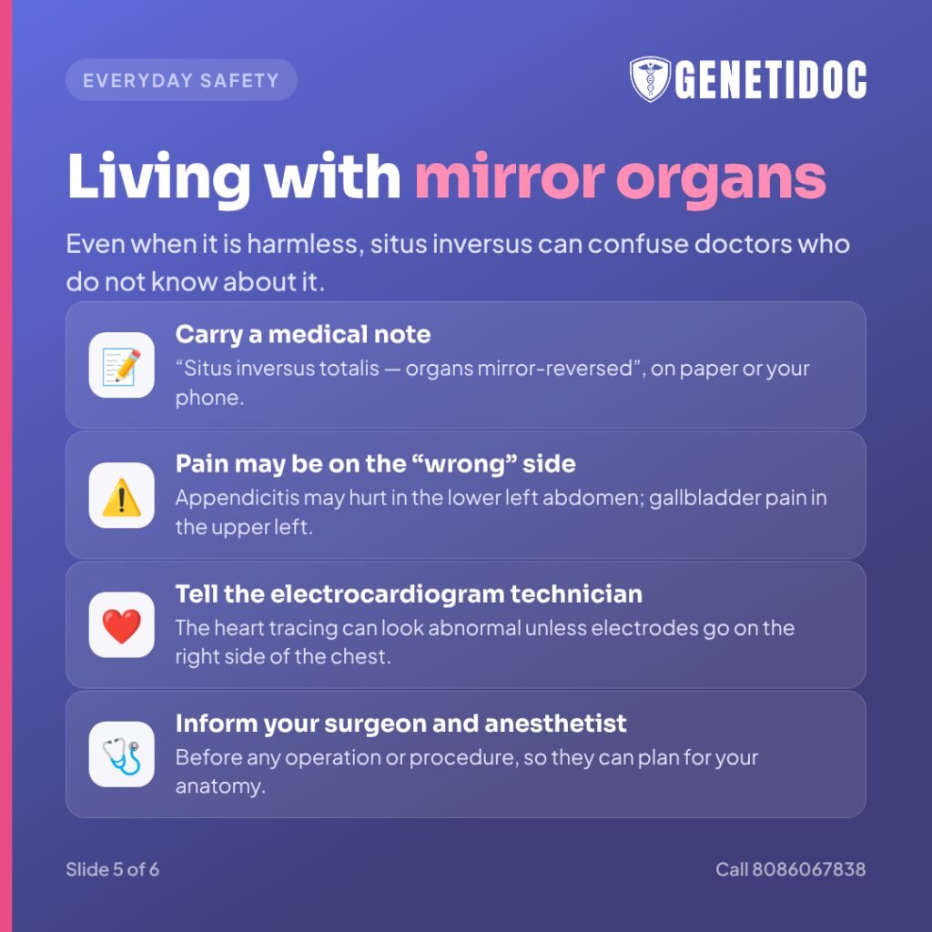
Even when situs inversus is harmless, it can confuse doctors who do not know about it. Practical steps help:
- Carry a medical note or keep it on your phone, stating “situs inversus totalis — organs mirror-reversed.”
- Know that pain may appear on the “wrong” side. Appendicitis may cause pain in the lower left abdomen, and gallbladder pain may appear on the upper left side. Tell emergency doctors about your situs inversus straight away.
- Inform ECG technicians. A standard ECG can look abnormal unless electrodes are placed on the right side of the chest.
- Tell your surgeon and anesthetist before any operation or procedure, so they can plan for your anatomy.
Situs Inversus Found on a Pregnancy Scan
Abnormal organ position is sometimes first seen on the routine anomaly scan, usually done around 18 to 20 weeks of pregnancy. This can be frightening, but the outlook depends heavily on exactly what is seen.
What usually happens next
- A detailed fetal heart scan (fetal echocardiography) to check the heart’s structure carefully
- A detailed review by a fetal medicine specialist to map the organs and look for other findings
- Genetic counseling to explain the possibilities, the testing options and what results can and cannot tell you
- Discussion of diagnostic testing, such as amniocentesis with chromosomal microarray, where appropriate. This is arranged through a fetal medicine specialist.
What parents should know
- A baby with complete mirror-image organs and a normal heart usually has a good outlook. Many grow up healthy.
- A baby with heterotaxy and a complex heart defect needs a delivery plan at a center with pediatric cardiac care.
- Whether a baby has PCD usually cannot be confirmed from a scan. Newborns with abnormal organ position should be watched closely for breathing difficulty after birth and evaluated if symptoms appear.
- Non-Invasive Prenatal Testing (NIPT), the blood test for chromosome conditions, does not detect situs inversus, heterotaxy or PCD.
Family Planning and Recurrence Risk
Once a definite genetic diagnosis is made in one family member, it opens several options:
- Testing brothers and sisters. Siblings of a child with PCD may also have PCD, sometimes with normal organ position and milder symptoms that have been overlooked.
- Carrier testing for relatives and their partners, especially in families with marriages between relatives
- Prenatal diagnosis in future pregnancies, through a fetal medicine specialist
- Preimplantation genetic testing with IVF, so that embryos without the condition can be selected
These options rely on knowing the exact disease-causing genetic change. This is one more reason why a clear, well-interpreted result in the first affected person is so valuable. An uncertain result cannot be used for these decisions.
Fertility and PCD
Sperm swim using a tail called a flagellum, which is built much like a cilium. In many men with PCD, sperm cannot move properly, causing infertility. In women, cilia in the fallopian tubes help move the egg, so some women with PCD have difficulty conceiving or a higher risk of ectopic pregnancy.
Infertility in PCD is very often treatable. Assisted reproduction techniques, particularly ICSI (intracytoplasmic sperm injection, where a single sperm is injected directly into an egg), can help many men father children. Genetic counseling before treatment helps couples understand the chance of passing the condition on, and whether testing the partner as a carrier makes sense.
Illustrative Examples
The following are composite scenarios based on typical presentations described in the medical literature. They are not specific Genetidoc patients.
Example 1: “Just a curiosity” — for 30 years
A man in his thirties has known since childhood that his heart is on the right. He has also had a wet cough and blocked nose for as long as he can remember, and repeated courses of antibiotics. When he and his wife cannot conceive, a semen test shows sperm that barely move. Putting the pieces together — mirror-image organs, lifelong chest and sinus symptoms, and infertility — points to PCD. Genetic testing confirms it. He starts regular airway clearance, and the couple is referred for assisted reproduction with genetic counseling.
Example 2: The sibling nobody suspected
A child with mirror-image organs and recurrent pneumonia is diagnosed with PCD. Both parents, who are cousins, are found to be carriers. Her older brother has normal organ position but has always had “asthma” that never fully responds to inhalers. Targeted testing for the family’s genetic changes shows that he also has PCD. Without the sister’s diagnosis, his condition might have gone unrecognized for years.
Example 3: Reassurance on a pregnancy scan
An anomaly scan shows the baby’s heart and stomach on the right side. A detailed fetal heart scan finds a structurally normal heart with all organs neatly mirrored. After counseling, the parents understand that the outlook is generally good. They also know to watch for breathing difficulty after birth, and that the baby can be evaluated for PCD if symptoms such as a persistent cough appear.
Common Misconceptions
- “Situs inversus is always harmless.” Complete, isolated situs inversus often is. But it can also be the first clue to PCD or heterotaxy.
- “Normal organ position rules out PCD.” Not true. About half of people with PCD have organs in the normal position.
- “Something during pregnancy caused this.” In most cases, nothing the mother did or did not do caused it. Organ position is set in the very first weeks of development, often before a woman knows she is pregnant.
- “My child’s cough is just allergy.” A wet, daily cough all year round since infancy is not typical allergy and deserves investigation.
- “Only the affected child needs attention.” Brothers, sisters and future pregnancies may be affected too, which makes a clear family diagnosis important.
- “A negative genetic test means it is not genetic.” A negative result means nothing reportable was found in what was tested. It is not a guarantee.
“One diagnosis in a family often helps more than one person. When we find the cause in one child, we can check siblings, guide relatives, and give parents real choices for the next pregnancy.”
— Dr. Roshan Daniel, Clinical Geneticist, Genetidoc
What Should You Do Next? A Practical Checklist
- Get a heart check. Have an echocardiogram if you have not had one.
- Look honestly at the symptoms. Is there a lifelong wet cough, blocked nose, ear problems, breathing trouble at birth, or fertility difficulty?
- Write down your family history. Note relatives with similar problems, early childhood deaths, heart defects, infertility, and whether the parents are related.
- Gather your reports. Bring X-rays, scans, CT reports, echocardiograms and any previous genetic reports.
- See a clinical geneticist if any red flags are present, or if you are planning a family.
- Keep a medical note about your situs inversus with you at all times.

Frequently Asked Questions
1. What is situs inversus?
Situs inversus is a condition present from birth in which the chest and abdominal organs are arranged as a mirror image of the usual layout. The heart is on the right, the liver on the left, and the stomach on the right.
2. Is situs inversus dangerous?
Complete, isolated situs inversus is usually not dangerous and does not shorten life. It needs closer evaluation if it is linked to primary ciliary dyskinesia, heterotaxy or a heart defect.
3. What is the life expectancy of someone with situs inversus?
People with isolated situs inversus totalis generally have a normal life expectancy. Outlook in PCD or heterotaxy depends on lung health, heart defects and the quality of ongoing care.
4. Is situs inversus genetic?
It can be. When it is part of PCD or heterotaxy, a genetic cause can often be found. PCD is usually inherited when both parents are healthy carriers. Isolated cases often have no identifiable cause.
5. What is the difference between situs inversus and dextrocardia?
Dextrocardia means the heart is on the right side of the chest. Situs inversus totalis means all the organs are reversed, including the heart. Dextrocardia can also occur on its own, which is more often linked to heart defects.
6. What is Kartagener syndrome?
Kartagener syndrome is the combination of primary ciliary dyskinesia with situs inversus. It causes chronic sinus and chest infections, bronchiectasis and often infertility, alongside mirror-image organs.
7. Can people with situs inversus have children?
Yes. People with isolated situs inversus usually have normal fertility. Those with PCD may face infertility, but assisted reproduction such as ICSI often helps.
8. Can situs inversus be detected before birth?
Yes. Abnormal organ position can often be seen on the anomaly scan. A fetal heart scan and genetic counseling help clarify the outlook. NIPT does not detect it.
9. If one child has PCD, what is the risk for the next pregnancy?
When both parents are carriers of autosomal recessive PCD, each pregnancy has a 1 in 4 chance of an affected child. Prenatal diagnosis or preimplantation genetic testing can be offered once the family’s genetic changes are confirmed.
10. Where would appendicitis pain be in someone with situs inversus?
Because the appendix is on the left, appendicitis pain is often felt in the lower left abdomen. Always tell emergency doctors about your situs inversus.
11. Which genetic test is used for situs inversus?
It depends on the clinical picture. Options include a PCD gene panel, a heterotaxy panel, chromosomal microarray, whole exome sequencing, or targeted testing if the family’s variant is already known.
12. Does marriage between relatives increase the risk?
Yes, for recessive conditions like PCD. Related parents are more likely to carry the same rare genetic change. Genetic counseling and carrier testing can help families understand their risk.
Key Takeaways
- Situs inversus means mirror-reversed organs. On its own, it is usually harmless.
- It can be the first clue to primary ciliary dyskinesia or heterotaxy, which need specialist care.
- A lifelong wet cough, a blocked nose, breathing trouble at birth, ear problems or infertility alongside mirror-image organs should prompt evaluation.
- Choosing the right genetic test, and interpreting it carefully, is essential. This is especially true in India, where population-specific genetic changes and consanguinity matter.
- A clear diagnosis improves lung and heart care, protects siblings and supports informed family planning.
Mirror-Image Organs and a Cough That Never Goes Away? Let’s Find Out Why.
If you or your child has situs inversus, dextrocardia or heterotaxy, especially with chest, sinus, ear or fertility problems, or if a pregnancy scan has shown abnormal organ position, our clinical geneticists can help. We review your scans and history, choose the right test for your situation, and explain what the results mean for you and your family.
Consult in person at our Thiruvananthapuram clinic or by teleconsultation from anywhere in India, with counseling available in 15+ languages.
Book a genetic consultation for situs inversus and primary ciliary dyskinesia
Have a question about organ position, PCD or heterotaxy? Join the conversation on the genetidoc rare genetic diseases forum
Related Reading
- Genetic Causes of Recurrent IVF Failure: What Couples in India Should Know Before Another Cycle
- Genetics and Obesity: Is It All in the Genes?
- Hypertrichosis: The Genetics Behind Excess Hair Growth
- Migraine and Genetics: Is It Hereditary?
- Mucopolysaccharidosis: Signs Parents Should Not Ignore
References
- Zariwala MA, Knowles MR, Leigh MW. Primary Ciliary Dyskinesia. In: Adam MP, et al., editors. GeneReviews®. University of Washington, Seattle. https://www.ncbi.nlm.nih.gov/books/NBK1122/
- Shapiro AJ, Davis SD, Polineni D, et al. Diagnosis of Primary Ciliary Dyskinesia. An Official American Thoracic Society Clinical Practice Guideline. American Journal of Respiratory and Critical Care Medicine. 2018;197(12):e24–e39.
- Lucas JS, Barbato A, Collins SA, et al. European Respiratory Society guidelines for the diagnosis of primary ciliary dyskinesia. European Respiratory Journal. 2017;49(1):1601090.
- Kennedy MP, Omran H, Leigh MW, et al. Congenital heart disease and other heterotaxic defects in a large cohort of patients with primary ciliary dyskinesia. Circulation. 2007;115(22):2814–2821.
- Shapiro AJ, et al. Laterality Defects in Primary Ciliary Dyskinesia: Relationship to Ultrastructural Defect or Genotype. Annals of the American Thoracic Society. 2023;20(3):397–405. doi:10.1513/AnnalsATS.202206-487OC
- Rosenfeld M, Ostrowski LE, Zariwala MA. Primary ciliary dyskinesia: keep it on your radar. PMC6040643.
- Gatt D, Golan Tripto I, Levanon E, et al. Stepwise genetic approach for the diagnosis of primary ciliary dyskinesia in highly consanguineous populations. Archives of Disease in Childhood. 2024;109(5):428.
- Genetic profile of children with suspected primary ciliary dyskinesia from India: a prospective cross-sectional whole exome sequencing study (162 children). [Full author/journal citation to be verified before publishing.]
- Cowan JR, et al. Copy number variation as a genetic basis for heterotaxy and heterotaxy-spectrum congenital heart defects. Philosophical Transactions of the Royal Society B. 2016. PMC5104505.
- Rafie NNA, Yubbu P, Aissvarya S, et al. Genetic aspects of congenital heart disease in heterotaxy syndrome. Revista Portuguesa de Cardiologia. 2025. doi:10.1016/j.repc.2025.05.007
- MedlinePlus Genetics. Primary ciliary dyskinesia. National Library of Medicine. https://medlineplus.gov/genetics/condition/primary-ciliary-dyskinesia/
- ARUP Laboratories. Heterotaxy and Situs Inversus Panel — Test Fact Sheet (Epidemiology). 2023.
This article is for educational purposes and does not replace a consultation with a qualified doctor. Decisions about testing and treatment should be made with your healthcare team.