A baby whose eyes do not seem to follow faces. A toddler who keeps bumping into furniture at dusk. A white glow in the pupil that appears only in a flash photograph. A school-age child whose glasses never seem strong enough.
Most childhood vision problems, such as simple short-sightedness or a mild squint, are common and easily treated. But some have a genetic cause. These conditions are not rare in India, and finding the exact cause early can change how a child is treated, which other organs are checked, and what the family knows about future children.
This guide explains which childhood eye conditions can be genetic, the warning signs parents should not ignore, how genetic testing works, and what a diagnosis actually changes for your child and your family.
Quick answer: What is a genetic eye disease?
A genetic eye disease is a vision problem caused by a change in one or more genes that control how the eye develops or works. In children, the most common examples are inherited retinal dystrophies (such as retinitis pigmentosa and Leber congenital amaurosis), congenital cataract, congenital glaucoma, albinism, aniridia and retinoblastoma. Some affect only the eyes. Others are the first sign of a condition that also affects hearing, the kidneys, the brain or metabolism.
How Common Are Genetic Eye Conditions in Indian Children?
Inherited eye disorders are an important cause of visual impairment in children worldwide. In India, several factors make them especially relevant:
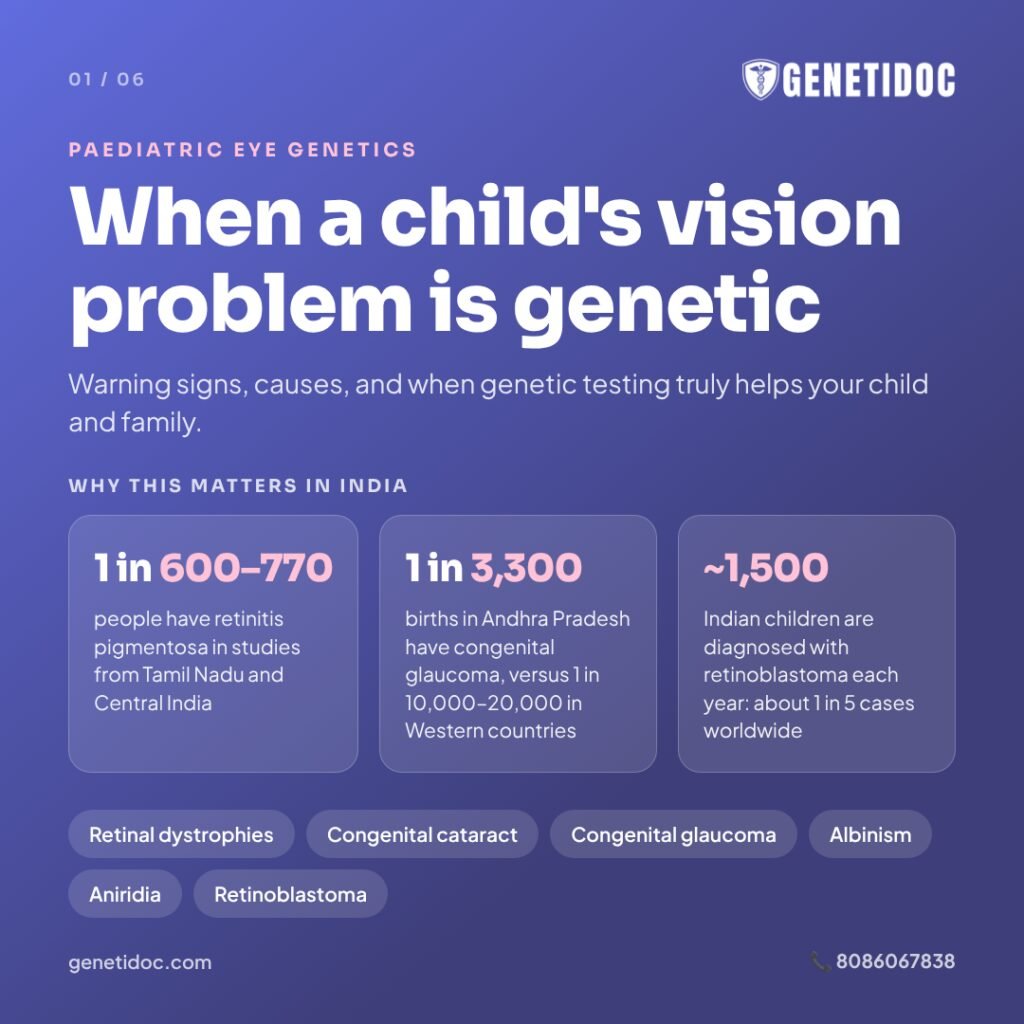
- Retinitis pigmentosa, the most common inherited retinal condition, has been found in about 1 in 600 to 1 in 770 people in population studies from Tamil Nadu and Central India. It is often linked to marriages between blood relatives in some South Asian communities.
- Primary congenital glaucoma has been reported in about 1 in 3,300 births in Andhra Pradesh, compared with roughly 1 in 10,000 to 20,000 in Western countries.
- Retinoblastoma, a childhood eye cancer, is diagnosed in an estimated 1,500 Indian children every year. India carries about one in five of all retinoblastoma cases in the world.
Consanguineous marriage, where partners are related by blood, increases the chance that both parents carry the same silent gene change. This makes recessive eye conditions more common in some communities.
Warning Signs: When Should Parents Suspect a Genetic Eye Problem?
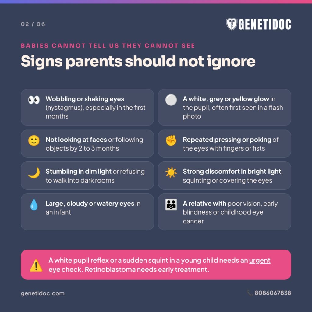
Babies cannot tell us they cannot see well. These signs deserve a prompt examination by a paediatric ophthalmologist (a children’s eye specialist):
- Wobbling or shaking eyes (nystagmus), especially if noticed in the first few months of life.
- A white, grey or yellow reflection in the pupil (leukocoria), often first seen in a flash photograph instead of the normal red-eye.
- Poor visual attention: a baby who does not look at faces or follow objects by 2 to 3 months.
- Repeated pressing or poking of the eyes with fingers or fists.
- Difficulty seeing in dim light or at night, such as stumbling in the evening or refusing to walk in dark rooms.
- Strong discomfort in bright light, with squinting or covering the eyes outdoors.
- Large, cloudy or watery eyes in an infant, which can be a sign of congenital glaucoma.
- A cloudy lens (cataract) or a missing-looking iris, noticed by the parents or a doctor.
- Very high or rapidly worsening short-sightedness in a young child.
- Another family member with a similar eye problem, early blindness, or a childhood eye cancer.
A white pupil reflex or a sudden squint in a young child should be checked urgently, because retinoblastoma needs early treatment.
The Main Genetic Eye Conditions in Children
Over 400 genes have been linked to inherited eye disorders. The table below gives an overview of the conditions most often seen in children.
| Condition | Part of the eye affected | Typical early signs | Examples of genes |
| Inherited retinal dystrophies | Retina (light-sensing layer) | Nystagmus, night blindness, light sensitivity, eye poking | RPE65, CEP290, GUCY2D, ABCA4, RPGR, USH2A |
| Congenital cataract | Lens | White pupil, poor visual attention, nystagmus | Crystallin genes, GJA8, OCRL, GALT |
| Primary congenital glaucoma | Drainage angle of the eye | Large, cloudy, watery eyes, light sensitivity | CYP1B1, LTBP2 |
| Albinism | Pigment in iris and retina; nerve pathways | Nystagmus, light sensitivity, pale iris; skin and hair may or may not be pale | TYR, OCA2, GPR143 |
| Aniridia | Iris and whole eye | Absent or partial iris, nystagmus | PAX6 (with WT1 in WAGR syndrome) |
| Retinoblastoma | Retina (cancer) | White pupil reflex, new squint | RB1 |
| Lens dislocation (ectopia lentis) | Lens position | High, changing short-sightedness | FBN1, CBS, ADAMTSL4 |
Inherited retinal dystrophies
The retina is the thin, light-sensing layer at the back of the eye. Inherited retinal dystrophies (IRDs) are a large group of conditions in which the retina’s light-sensing cells do not work properly or slowly wear out.
- Leber congenital amaurosis causes severe vision loss from birth or early infancy. Babies often have nystagmus, poor visual attention and a habit of pressing on their eyes.
- Retinitis pigmentosa usually begins with night blindness and gradual loss of side vision. It can start in childhood or later.
- Stargardt disease affects central vision, often noticed when a school-age child struggles to read the board.
- Achromatopsia causes poor colour vision, strong light sensitivity and reduced sharpness of vision from infancy, but usually stays stable.
In its 2025 teaching monograph, the All India Ophthalmological Society notes that the diagnosis of an IRD is incomplete without genetic testing, because conditions that look alike can have very different causes and outlooks.
Congenital and childhood cataract
A cataract is a clouding of the lens, which is normally clear. In a baby, a dense cataract blocks the image from reaching the retina just when the brain is learning to see. Without timely surgery, this can lead to permanent poor vision called amblyopia.
Many cataracts in both eyes, with no other cause found, are genetic. Some are part of wider conditions:
- Galactosemia: a metabolic condition in which the body cannot process galactose, a sugar in milk. Early cataracts can improve once a galactose-free diet is started.
- Lowe syndrome: a condition passed down on the X chromosome that mainly affects boys, and also affects the kidneys and development.
- Chromosomal conditions such as Down syndrome.
Infections in pregnancy, such as rubella, can also cause cataracts, so not every congenital cataract is genetic.
Primary congenital glaucoma
In this condition, the eye’s internal drainage system does not form properly. Pressure builds up inside the eye, which stretches it and damages the optic nerve. Affected babies often have large, cloudy, watery eyes and dislike light.
In Indian studies, changes in the CYP1B1 gene were found in about 1 in 3 children with primary congenital glaucoma. It is usually inherited in a recessive pattern, meaning both parents silently carry one changed copy.
Albinism
Albinism reduces the pigment melanin in the eyes, and often in the skin and hair too. In the eye, this affects how the centre of the retina and the nerve pathways develop. Children usually have nystagmus, light sensitivity and reduced vision.
Some children have only the eye form, called ocular albinism, with normal-looking skin and hair for their family. This can make albinism easy to miss without careful examination or genetic testing.
Aniridia
Aniridia means the coloured part of the eye (the iris) is partly or completely missing. It is caused by changes in the PAX6 gene and affects the whole eye, with a lifelong risk of glaucoma, cataract and corneal problems.
Aniridia is a clear example of why genetic testing matters beyond the eye. In some children with no family history, the gene deletion also includes a nearby gene called WT1. This causes WAGR syndrome, named after its main features (Wilms tumour, aniridia, genital and urinary differences, and developmental delay). Children with WAGR syndrome have a high risk of Wilms tumour, a childhood kidney cancer. Children with this deletion need regular kidney ultrasound scans, usually every three months, until about age eight. Children whose testing shows no WT1 deletion can be spared these scans.
Retinoblastoma
Retinoblastoma is a cancer of the retina that usually appears before age five. It is caused by changes in both copies of the RB1 gene inside retinal cells.
- In the heritable form, a child is born with one changed copy of RB1 in every cell of the body. Tumours are often in both eyes, and the child’s brothers, sisters and future children can be at risk.
- In the non-heritable form, both changes happen only in one eye’s tumour. The risk to relatives is low.
All children with retinoblastoma in both eyes, and some with it in one eye, have the heritable form. Genetic testing is the most reliable way to tell the two apart.
Why the eye is the first clue to a wider condition
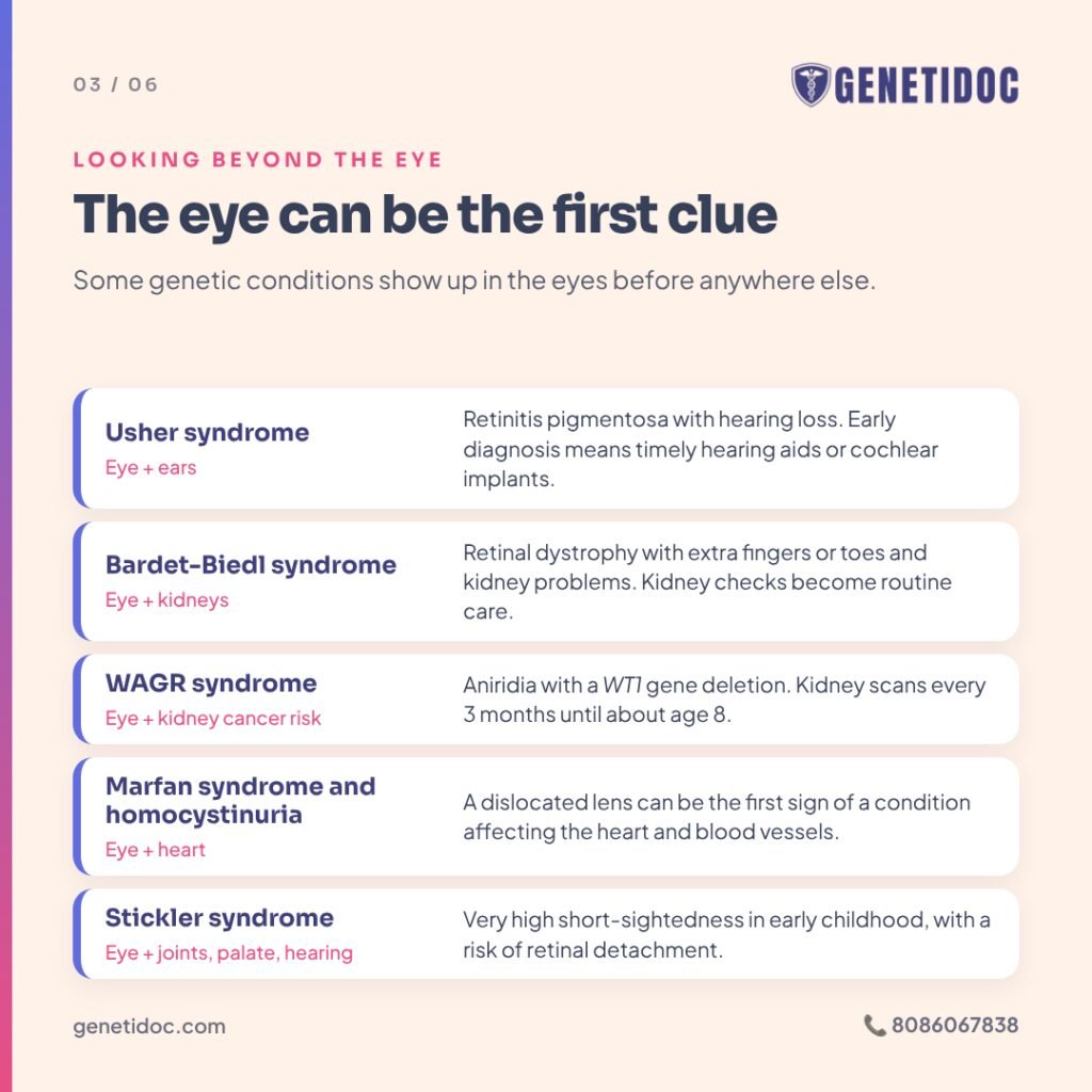
Some genetic conditions show up in the eyes first, before other features become obvious. Recognising them early changes a child’s care:
- Usher syndrome: retinitis pigmentosa together with hearing loss. Early diagnosis supports timely hearing aids or cochlear implants and planning for schooling.
- Bardet-Biedl syndrome: retinal dystrophy with extra fingers or toes, weight gain, kidney problems and learning difficulties. Kidney checks become part of routine care.
- Joubert syndrome and related ciliopathies: retinal and eye movement problems with brain, kidney and liver involvement.
- Marfan syndrome and homocystinuria: a dislocated lens can be the first sign of a condition that also affects the heart and blood vessels.
- Stickler syndrome: very high short-sightedness in early childhood, with a risk of retinal detachment, cleft palate, joint problems and hearing loss.
Two illustrative situations
The following examples are illustrative scenarios, not real patients.
The baby with wobbling eyes. Consider a 4-month-old whose parents notice her eyes shaking side to side. She does not follow toys well and often presses her fists into her eyes. Her parents are first cousins. The eye specialist suspects Leber congenital amaurosis, and an electroretinogram (ERG), a test that measures the retina’s electrical response to light, shows almost no response. A retinal gene panel finds changes in both copies of one gene. The family now knows the exact diagnosis, whether any treatment or research trial applies, and that each future pregnancy has a 1 in 4 chance of being affected.
The boy with night blindness and poor hearing. Now consider a 9-year-old who has struggled in school, wears hearing aids, and has recently started tripping on the stairs after sunset. His family assumed the two problems were unrelated. An eye examination shows early retinitis pigmentosa, and genetic testing confirms Usher syndrome. His school support, hearing care and eye follow-up can now be planned together.
How Genetic Testing Works for Childhood Eye Conditions
Genetic testing is done on a small blood or saliva sample. It works best as part of a step-by-step process, not as a standalone test.
- A detailed eye examination by a paediatric ophthalmologist, sometimes under anaesthesia in young children.
- Special eye tests where needed, such as an ERG or retinal imaging, to show which part of the eye is affected.
- A full assessment of the child by a clinical geneticist, looking for features outside the eye such as hearing, kidneys, development, growth and facial features.
- A three-generation family history, including whether the parents are related by blood.
- Pre-test genetic counseling, so the family understands what the test can and cannot show.
- Choosing the right test, based on all of the above.
- Interpretation and post-test counseling, linking the result to the child’s care and the family’s plans.
Which test is chosen?
At Genetidoc, the scope of testing is matched to how clearly the child’s features point to a known condition.
- Single-gene testing: when one gene is strongly suspected, such as RB1 in retinoblastoma or PAX6 in aniridia. Deletion testing is added where needed, because some changes are too large for routine sequencing to detect reliably.
- Targeted family-variant testing: when the gene change in the family is already known. Results usually take about 1 week.
- Eye disease gene panels: for conditions with many possible genes, such as retinal dystrophies or congenital cataract. Results usually take about 3 to 4 weeks.
- Whole exome sequencing: when the eye problem is part of a wider picture involving development, hearing, kidneys or other organs. Results usually take about 1 to 2 months.
- Chromosomal microarray: when a missing or extra piece of a chromosome is suspected, for example a child with aniridia and developmental delay.
- Whole genome sequencing: kept as a next step when a genetic condition is strongly suspected but earlier testing was negative or incomplete.
How often does testing find the cause?
Results from specialist centres are encouraging:
- In a UK study of 201 children aged 0 to 5 years with inherited eye disorders, genetic testing found the cause in 64% overall, ranging from 39% to 91% depending on the condition.
- In children with inherited retinal disease, multi-gene panel testing pointed to a genetic diagnosis in about 4 out of 5 children.
Testing works best when the eye findings are carefully described first. A well-chosen test in a well-examined child gives far more useful answers than a large test ordered without a clear clinical picture.
What if the result is negative or uncertain?
A negative result means no reportable change was found in the genes tested. It does not mean the condition is not genetic. Some genes are not yet known, and some types of changes are hard to detect with certain tests.
A variant of uncertain significance (VUS) is a gene change that science cannot yet classify as harmful or harmless. It is neither a positive nor a negative result.
- Care is guided by the eye examination, not by the uncertain variant.
- Testing affected relatives for the same variant can sometimes help clarify it.
- A VUS is not used to test healthy relatives or to make pregnancy decisions.
- Results can be reanalysed later, usually after 6 months to 1 year, or sooner if new symptoms appear. Keep your report and ask for a copy of the raw data.
What Does a Genetic Diagnosis Actually Change?
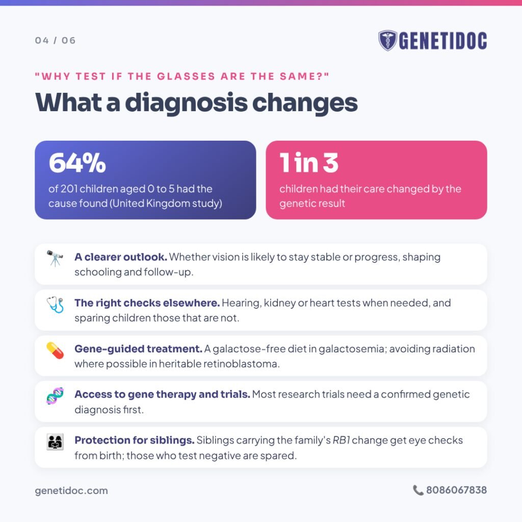
Parents sometimes ask, “If glasses and surgery are the same either way, why test?” In the UK study above, the genetic result changed the child’s management in about one in three cases. It also avoided unnecessary tests and allowed families to plan ahead.
1. A clearer picture of the future
Two children can have the same symptoms at age two but very different futures. Genetic testing can help show whether a retinal condition is likely to stay stable or slowly progress. This shapes schooling, career guidance and how closely the eyes are followed.
2. Checking the rest of the body
A diagnosis can trigger hearing tests in Usher syndrome, kidney checks in Bardet-Biedl syndrome or Lowe syndrome, kidney ultrasound scans in WAGR syndrome, and heart scans in Marfan syndrome. It can also rule these out, sparing children unnecessary investigations.
3. Treatment that depends on the gene
- In galactosemia, a galactose-free diet protects the eyes, liver and brain.
- In homocystinuria, treatment with vitamin B6, diet and other measures reduces complications, including the risk of blood clots around surgery and anaesthesia.
- In heritable retinoblastoma, doctors try to avoid radiation where possible, because these children have a higher lifelong risk of other cancers.
4. Gene therapy and clinical trials
The first approved gene therapy for an inherited eye disease, voretigene neparvovec (Luxturna), treats retinal dystrophy caused by changes in both copies of the RPE65 gene. It works only while enough living retinal cells remain, and only for this specific gene.
This therapy is not routinely available in India at present, and its cost is very high. Research trials for several other retinal genes are ongoing worldwide. Most require a confirmed genetic diagnosis before a child can even be considered. Knowing the exact gene keeps that door open.
5. Protecting brothers, sisters and future children
Once the gene change is known, relatives can be offered targeted testing for that single change. In retinoblastoma, this is especially important. Brothers and sisters who carry the family’s RB1 change need regular eye examinations from birth, often under anaesthesia, so any tumour is found while it is small. Those who test negative can be spared years of repeated examinations.
How Are Genetic Eye Conditions Inherited?
Understanding the inheritance pattern helps families understand the chance of it happening again.
- Autosomal recessive: both parents are healthy carriers. Each pregnancy has a 1 in 4 (25%) chance of an affected child. This is the most common pattern in childhood retinal dystrophies and congenital glaucoma, and is more likely when parents are related by blood.
- Autosomal dominant: one changed copy is enough. An affected parent has a 1 in 2 (50%) chance of passing it on. Many isolated congenital cataracts and aniridia follow this pattern.
- X-linked: the gene is on the X chromosome. Boys are usually more severely affected, and mothers may be carriers with mild or no symptoms. Examples include some forms of retinitis pigmentosa, ocular albinism and Lowe syndrome.
- New (de novo) changes: the gene change starts in the child and is not carried by either parent. The chance of recurrence is usually low, but not zero.
A condition that “skipped a generation” or appeared with no family history can still be genetic. This is one of the most common misunderstandings families have.
Planning future pregnancies
When the gene change in the family is known, couples have options. These include testing in a future pregnancy through chorionic villus sampling or amniocentesis, or preimplantation genetic testing during in vitro fertilisation (IVF) to select embryos without the family’s gene change. These are deeply personal choices. Genetic counseling helps couples understand each option, and the decision always rests with them.
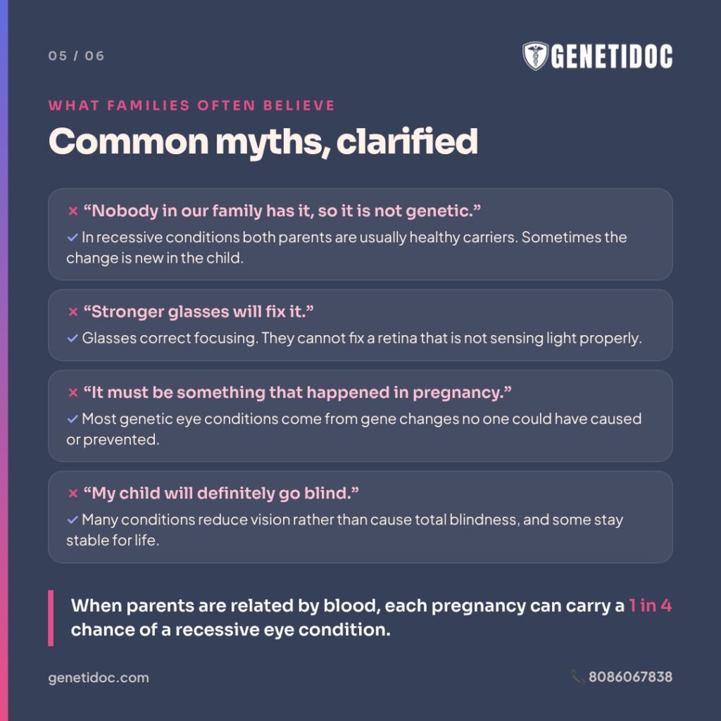
Common Misconceptions About Childhood Eye Conditions
“Stronger glasses will fix it.”
Glasses correct focusing problems. They cannot fix a retina that is not sensing light properly. If vision stays poor despite the right glasses, the cause needs to be found.
“It must be something that happened during pregnancy.”
Parents, especially mothers, often blame themselves. Most genetic eye conditions are caused by gene changes that no one could have caused or prevented.
“Nobody else in our family has it, so it is not genetic.”
In recessive conditions, both parents are usually healthy carriers. In other cases, the change is new in the child. A family history is not needed for a condition to be genetic.
“Nothing can be done, so why test?”
Even without a cure, a diagnosis guides checks of other organs, avoids unnecessary tests, informs schooling and career planning, protects relatives, and opens the door to research trials.
“My child will definitely go blind.”
Many genetic eye conditions cause reduced vision, not total blindness, and some remain stable for life. The exact diagnosis gives a far more accurate picture than general fears.
When Genetic Testing May Not Be Needed
- Common refractive errors, such as ordinary short-sightedness, long-sightedness or astigmatism, without other worrying features.
- A simple squint in an otherwise healthy child with normal eye examination findings.
- A cataract clearly caused by injury, infection or medicines.
- Retinopathy of prematurity, which is linked to premature birth rather than an inherited gene change.
- Healthy children with no symptoms, when the family condition is untreatable and adult-onset, except where knowing early helps the child, as in retinoblastoma.
The American Academy of Ophthalmology (AAO) also advises against direct-to-consumer genetic tests for eye disease, and against broad testing that is not guided by a clinical diagnosis.
Why Expert Interpretation Matters
- Choosing the right test. A small retinal panel can miss a syndrome. A test that only looks for small changes can miss large deletions, such as those in aniridia or retinoblastoma.
- Reading the result correctly. Indian populations are under-represented in global genetic databases, so new and uncertain variants are common. Each result needs careful review against the child’s actual eye findings.
- Looking beyond the eye. A clinical geneticist checks whether the result explains the whole child and which other specialists should be involved.
- Counseling the family. Professional guidance from the AAO states that genetic counseling should be offered to everyone who has genetic testing, because results can affect family planning, relationships and feelings of guilt.
At Genetidoc, testing is carried out through accredited partner laboratories. Every result is reviewed by a clinical geneticist and explained through genetic counseling. Families then return to their paediatric ophthalmologist and other specialists with a clear, written plan.
Living Well With a Genetic Eye Condition
- Start early intervention. Babies and toddlers with low vision benefit from early stimulation and developmental support.
- Use low-vision aids. Magnifiers, large-print materials, tinted glasses for light sensitivity and screen-reading technology can make a big difference at school.
- Talk to the school. Seating near the board, extra time in exams and accessible materials help children reach their potential.
- Know your entitlements. In India, blindness and low vision are recognised disabilities under the Rights of Persons with Disabilities Act, 2016. Eligible children can apply for a disability certificate and a Unique Disability ID (UDID) card, which can help with educational and other support.
- Protect the eyes. Sunglasses and hats help children with albinism and light sensitivity. Children with very high short-sightedness should know the warning signs of retinal detachment, such as sudden flashes, floaters or a shadow over vision.
- Keep up regular follow-up, even when vision seems stable.
What To Do If You Are Worried About Your Child’s Vision
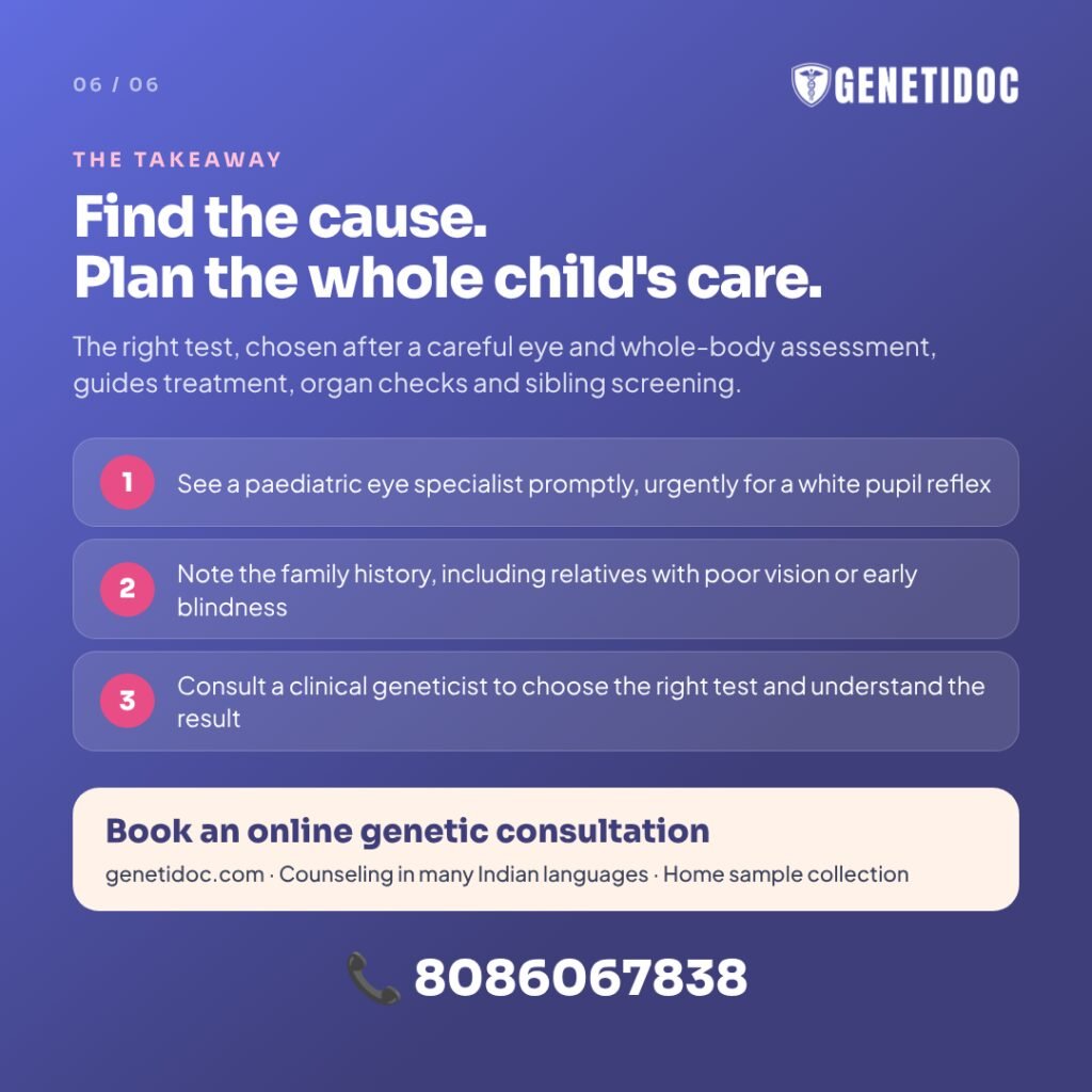
- See a paediatric ophthalmologist promptly, and urgently if you see a white pupil reflex.
- Take photos and videos of the eye movements or the pupil reflex to show the doctor.
- Write down the family history, including any relatives with poor vision, early blindness, eye cancer, hearing loss, or parents related by blood.
- Ask whether the condition could be genetic, especially if both eyes are affected, symptoms began in infancy, or other health problems are present.
- Consult a clinical geneticist to choose the right test and understand what it means for your child and family.
- Keep every report, and return for review if new symptoms develop.
Frequently Asked Questions
1. Can eye problems in children be genetic?
Yes. Many childhood vision problems, including retinal dystrophies, congenital cataract, congenital glaucoma, albinism, aniridia and retinoblastoma, can be caused by gene changes.
2. What are the early signs of a genetic eye disease in a baby?
Wobbling eyes, not looking at faces, eye poking, a white pupil reflex, large cloudy eyes, strong light sensitivity or poor vision in dim light.
3. Is nystagmus in babies always genetic?
No, but it is often a sign of an underlying eye or brain condition, and many of these are genetic. Every baby with nystagmus should have a full eye examination.
4. Which genetic test is done for inherited eye disease?
It depends on the condition. Options include single-gene testing, eye disease gene panels, whole exome sequencing and chromosomal microarray. A clinical geneticist chooses the test based on the eye findings and the whole child.
5. How long do genetic test results take?
Targeted family-variant tests usually take about 1 week, eye gene panels about 3 to 4 weeks, and whole exome sequencing about 1 to 2 months.
6. Is there a treatment for retinitis pigmentosa or Leber congenital amaurosis?
Gene therapy is approved for retinal dystrophy caused by the RPE65 gene, but it is not routinely available in India. Trials for other genes are ongoing. Supportive care and low-vision aids help all children.
7. Can cousin marriage cause eye problems in children?
Marriage between blood relatives increases the chance that both parents carry the same recessive gene change. This raises the risk of conditions such as retinal dystrophies and congenital glaucoma.
8. Will my next child have the same eye condition?
It depends on the inheritance pattern. The risk may be 25% for recessive conditions, up to 50% for dominant conditions, and usually low for new changes. Genetic counseling gives your family’s specific risk.
9. Is retinoblastoma hereditary?
Some cases are. All children with tumours in both eyes, and some with tumours in one eye, have the heritable form. Genetic testing shows which relatives need regular eye screening.
10. Should my child’s brothers and sisters be tested?
If the gene change is known, targeted testing of siblings can be offered when it changes their care, such as in retinoblastoma or aniridia. Otherwise, a careful eye examination is a good first step.
11. What does a negative genetic test mean for an eye condition?
It means no reportable change was found in the genes tested. It does not rule out a genetic cause. The result can be reanalysed later as knowledge grows.
12. Can genetic testing be done on saliva instead of blood?
Some laboratories accept saliva samples. Blood is still the most common sample, and Genetidoc arranges home sample collection.
Key Takeaways
- Many childhood vision problems have a genetic cause, and some are more common in India because of consanguinity.
- Wobbling eyes, a white pupil reflex, eye poking, night blindness and large cloudy eyes all need prompt eye examination.
- The eye can be the first clue to a condition affecting hearing, kidneys, heart or metabolism.
- Genetic testing finds the cause in a large share of children when the test is chosen after a careful eye and whole-body assessment.
- A diagnosis can guide checks of other organs, treatment, research trial access, and screening of siblings.
- Negative and uncertain results need expert interpretation and later review.
- Genetic counseling helps families understand recurrence risk and make informed choices.
Has Your Child’s Eye Specialist Raised the Possibility of an Inherited Eye Condition?
If your child has nystagmus, a retinal dystrophy, congenital cataract or glaucoma, aniridia, albinism or retinoblastoma, or if poor vision runs in your family, a consultation with a clinical geneticist can help identify the cause. It can also guide the right test, check for features beyond the eye, and help plan care for your child’s brothers, sisters and future pregnancies.
Genetidoc offers online genetic counseling across India, with home sample collection and counseling in many Indian languages.
Book a paediatric eye genetics consultation
Have questions about your child’s eye condition, or want to connect with other families? Visit the Genetidoc Rare Disease Forum.
Related Reading
- Facial Features and Genetic Syndromes: A Guide for Parents
- Is Genetic Testing Useful for Unexplained Infertility?
- Family History of Dementia: When to Seek Genetic Counseling
- APOE and Alzheimer’s Risk: Separating Fact from Fear
- GM1 Gangliosidosis: Why Early Diagnosis Matters
- What Defines a Rare Genetic Disease? A Clear Guide
References
- Lenassi E, Clayton-Smith J, Douzgou S, et al. Clinical utility of genetic testing in 201 preschool children with inherited eye disorders. Genet Med. 2020;22(4):745–751. doi:10.1038/s41436-019-0722-8
- Taylor RL, et al. Panel-based clinical genetic testing in 85 children with inherited retinal disease. Ophthalmology. 2017;124(7).
- Stone EM, Aldave AJ, Drack AV, et al. Recommendations for genetic testing of inherited eye diseases: report of the American Academy of Ophthalmology task force on genetic testing. Ophthalmology. 2012;119(11):2408–2410.
- Kannabiran C, Parameswarappa D, Jalali S. Genetics of inherited retinal diseases in understudied populations. Front Genet. 2022;13:858556.
- All India Ophthalmological Society, Academic and Research Committee. CME Series: Inherited Retinal Dystrophies. 2025.
- Reddy ABM, Kaur K, Mandal AK, et al. Mutation spectrum of the CYP1B1 gene in Indian primary congenital glaucoma patients. Mol Vis. 2004;10:696–702.
- Tanwar M, Dada T, Sihota R, et al. Mutation spectrum of CYP1B1 in North Indian congenital glaucoma patients. Mol Vis. 2009;15:1200–1209.
- Moosajee M, Hingorani M, Moore AT. PAX6-Related Aniridia. In: Adam MP, et al., eds. GeneReviews. Seattle (WA): University of Washington, Seattle. Available at: https://www.ncbi.nlm.nih.gov/books/NBK1360/
- Gupta H, et al. Retinoblastoma genetics screening and clinical management. BMC Med Genomics. 2021;14:188.
- Impact of RB1 gene screening from blood collected on a single day from 411 family members of 113 retinoblastoma survivors in India. Eye (Lond). 2024;38(8):1575–1580.
- European Medicines Agency. Luxturna (voretigene neparvovec): EPAR summary for the public. Available at: https://www.ema.europa.eu/en/medicines/human/EPAR/luxturna
This article is for educational purposes and does not replace personal medical advice. Please consult a qualified doctor or clinical geneticist about your child’s individual situation.